
Research

Single-Impulse Panoramic Photoacoustic Computed Tomography (SIP-PACT)

Super sensitivity and specificity: Integration of multiscale PAT and photoswitchable proteins

PACT-guided Microrobots For Targeted Navigation in Intestines In Vivo

Photoacoustic topography through an ergodic relay (PATER)
Label-free photoacoustic microscopy: super speed and super contrast
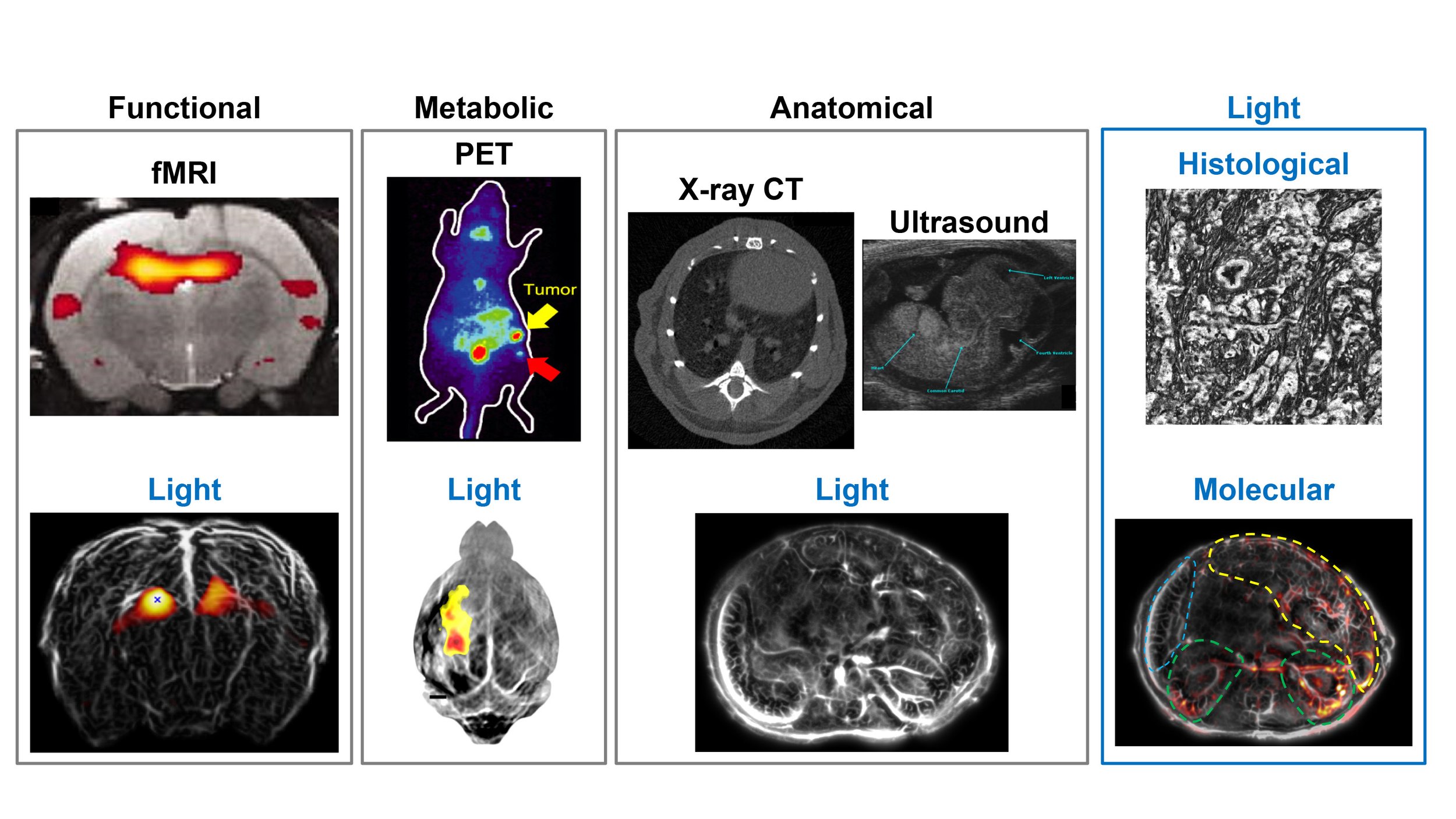
Why use light?
Fundamentally speaking, light interacts with tissue at the molecular level and allows us to probe all molecules directly. fMRI can provide functional information of the brain, so can light. PET can provide metabolic imaging, so does light. X-ray CT and ultrasound can provide anatomical imaging, so can light. In addition, light can also provide histological and molecular information. Moreover, unlike ionizing radiation used in x-ray CT or PET, it is very safe to use light. Photon energy is very gentle, only 2-3 eV in general, which is not high enough to ionize molecules.
Optical penetration
Biomedical optics have come a long way. The microscope used every day in modern medical diagnosis was invented in 17th-century, which revolutionized 19th-century medicine by letting us see into a cell. Then optical coherence tomography, developed in the 1990s, increased optical penetration to 1mm. Bringing huge benefits to clinical care for skin and eyes. Now photoacoustic imaging, first adopted for medical use in the 2000s, allows us to see even more, allowing penetration by another order of magnitude to several centimeters, allowing organ-level in vivo human imaging.

Optical imaging approaches visualize biological phenomena with sub-organelle resolutions at shallow depths. For clinical diagnosis, non-optical approaches, including magnetic resonance imaging (MRI), X-ray computed tomography (X-ray CT), and ultrasound tomography (UST), achieve excellent penetration. However, different modalities employ different imaging-contrast mechanisms, and each works within a certain spatial scale, hindering correlative perception of biological problems across scales. Moreover, the modalities suffer from certain limitations. For example, microscopic MRI is often too slow for imaging dynamics, and X-ray CT use ionizing radiation, inhibiting longitudinal observation. UST does not image blood oxygenation. As a notable exception, photoacoustic tomography (PAT) is capable of multiscale imaging, from organelles to small-animal whole bodies and human organs, using a consistent contrast of optical absorption.
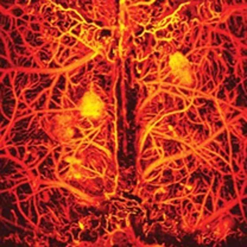
How PAT works?
We shine a gentle laser pulse onto the tissue, the light is absorbed, raising its temperature a bit. The rise in temperature leads to a tiny fraction of volume expansion, which in turn generates acoustic waves. Sensors process those sound signals, resulting in a high-resolution image that maps the optical absorption of the molecules in the biological tissue.